
Quick Review of Lisfranc Injuries
Lisfranc injures are a spectrum which result in a sprain or complete disruption of the tarsometatarsal joints of the midfoot. They most commonly occur at the base of the 2nd metatarsal with oftentimes subtle or even absent findings on standards x-ray views, especially when they result from low velocity injury.
What is mechanism of Injury?
Usually a result of plantar flexion with external rotation of the ankle (ie. fall from a horse with the foot caught in the foot stirrup, MVC, foot planted in a hole or step off a curb)
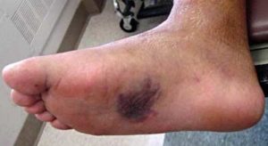
Physical Exam on Suspected Lisfranc Fracture
- Unable to bear weight
- Hematoma/ecchymosis on medial plantar aspect of foot
- Dorsal midfoot swelling
For suspected Lisfranc injuries you’ll need three views (AP, lateral and oblique). On normal foot x-rays you should notice alignment of the 2nd metatarsal on AP view and the medial edge of the base of the 2nd metatarsal should line up with the medial edge of the medial cuneiform. On the oblique view the 3rd and 4th metatarsal should have the medial edge of the 3rd and 4th metatarsal lining up with the medial edges of the middle and lateral cuneiform.
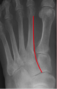
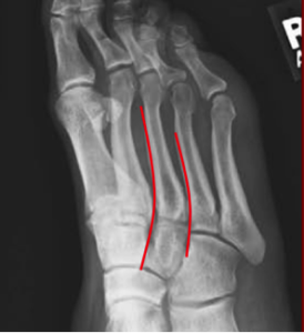
Common X-ray findings for Lisfranc Fracture.
- Widening of > 2mm between the base of the 1st and 2nd or 3rd and 4th metatarsal bases needs surgical intervention
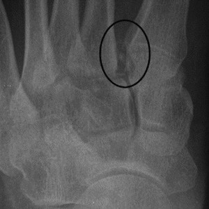
- “ Fleck Sign” is pathognomonic for a Lisfranc Injury. This is a small bony fragment avulsed from the 2nd metatarsal base or medial cuneiform
What if the X-ray is normal but a Lisfranc injury is still clinically suspected?
- Add a 30 degree oblique X-ray view
- Consider ankle nerve block with standing views (Weight bearing stress views). Similar to the example below with a normal appearing non weight bearing x-ray (left) and then weight-bearing (right) revealing the injury.
- In patients with clinically suspected Lisfranc injuries and normal or indeterminate radiographic findings, CT or MRI imaging is recommended.
- Given the superior depiction of soft tissue supporting structures and the ability of soft tissue supporting structures and the ability to detect soft tissue injuries in patients with unstable injuries on MR images, the American College of Radiology Appropriateness Criteria guidelines favors the use of MR imagining
- Orthopedics surgeons may request high resolution 3D CT images for preoperative planning and for depicting and further characterizing fractures.
ED management for patients with Lisfranc Injury
For nondisplaced or suspected injury without radiographic findings, you may place the patient in a posterior back slab. Patient should be non-weight bearing and arrange for orthopedic outpatient follow up two weeks later. For significantly displaced injury or dislocation (> 2mm widening at the Lisfranc Joint) , Immediate orthopedic referral is needed for urgent outpatient surgical intervention
Post by: Yenis Paez-Perez, DO