CC: Chest Pain
HPI: 49-year-old female brought in via ALS presents complaining of Chest pain. As per the Paramedics, the patient was found to be in no acute distress, stating she had exertional chest pain, which had subsided. The pre-hospital ECG was suspicious for ischemia and she was given ASA. Patient states she was walking home from the store when she began to have a pressure like pain on the left side of her chest, which was non-radiating and persisted when she laid down. She admits to feeling similar symptoms over the past few months, but today was the most severe. Upon arrival to Emergency Department, she denied chest pain, SOB, palpitations, abdominal pain, nausea or vomiting. Denied ETOH or illicit drugs use
Physical Exam:
BP:128/91, Hr:88, RR: 14, Temp: 98.0, Pulse O2: 100% RA
General: Patient lying comfortably in bed, in no distress
HEENT: NCAT, pupils PERRLA, neck supple
Respiratory: non-labored, CTA B/L, no wheezing, rales or rhonchi
Cardiac: +S1/S2, no MRG, regular rate and rhythm
Abdomen: soft NT ND, + BS
Neuro exam: AAO X 3, lucid, strength is 5/5 in all extremities, muscle tone is intact,
Skin: No rash or peripheral edema.
Pertinent Labs:
CBC: Unremarkable
CMP: Unremarkable
Troponin: Negative (< 0.010)
Pertinent Imaging/ECG
CXR: no acute infiltrate, No Pneumothorax or cardiomegaly. Normal Chest X-ray
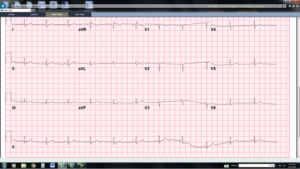
Pre-Hospital ECG

ECG in the ED

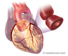
Working Diagnosis: Left Main Insufficiency
ED Course: Repeat ECG in ED showed NSR and had completely normalized. Case was discussed with Interventional cardiologist on call. A Code STEMI was activated and patient was taken emergently to the Cath lab. She was given a Heparin bolus as well as Plavix.
ED/Hospital course: The catheterization report revealed 80-90% Distal left main, 30 % mid LAD, and LM stenosis improved partially during catheterization with nitroglycerin. Patient admitted to recently using cocaine. CT surgery was called due to the fact that patient had recent cocaine use and it was believed she might have had Left Main Coronary spasm. On hospital day 2, patient went back for repeat catheterization, which revealed Left main distal 30% and LAD mid 30% stenosis. Patient was transferred to telemetry, and discharged on hospital day 4.
Pearls:
aVR in ACS
Typical ECG findings with left main coronary artery (LMCA) occlusion:
- Widespread horizontal ST depression, most prominent in Leads I, II and V4-6
- ST elevation in aVR ≥ 1mm
- ST elevation in aVR ≥ V1
(Don’t worry so much about STE 0.5mm or less in lead aVR, because it lacks specificity. Using 1.0mm or greater in lead aVR, has better specificity)
ST-Segment Elevation in lead aVR foreshadows a worse prognosis in ACS and often predicts the need for CABG. Patients with NSTEMI and ST elevation ≥ 1mm in aVR are likely to have multi-vessel or LMCA disease and are likely to require CABG, therefore withholding Clopidogrel may be prudent. ST-segment elevation in aVR can be caused by any of the following 4 mechanisms
- Critical narrowing of the LMCA causing sub-endocardial ischemia due to insufficient blood flow. (LMCA insufficiency)
- Transmural infarction of the basal septum due to a very proximal LAD occlusion or complete LMCA occlusion (patient will be VERY sick)
- Severe multi-vessel coronary artery disease.
- Diffuse sub-endocardial ischemia from oxygen supply/demand mismatch.
Patients with complete occlusion of the LMCA (mechanism 2), often present in cardiogenic shock and require immediate revascularization. Patients with acute coronary occlusions typically will have active symptoms and look sick!
There is an estimated 70% mortality without immediate PCI. Medical therapy (including thrombolytic) does not improve mortality. Emergency PCI may decrease mortality to 40%.
What Else can Cause STE in aVR that Won’t Benefit from Going to the Cath Lab?
Other causes of global cardiac ischemia
o Thoracic aortic dissection
o Large pulmonary embolism
o Severe anemia
o Post-arrest (within 15 min. of epinephrine or defibrillation)
oMiscellaneous causes
o Supraventricular Tachycardia (esp. AVRT)
o Left bundle branch block (LBBB) & paced rhythms
o LVH with strain (from severe hypertension)
o Severe hypokalemia
o Na+ channel blockade (TCA toxicity, hyperkalemia, Brugada, etc.)
REMEMBER: ST-Segment Elevation in Lead aVR is NOT SPECIFIC for an acute LMCA Lesion, Acute Proximal LAD Lesion, or Acute Triple Vessel DiseasE
-
- Correlate Your ECG with the Patient’s Clinical Status
- Patients with ACS due to LMCA Blockage, Triple Vessel Disease, or Proximal LAD blockage will look “sick” due to global cardiac ischemia
Case presented by Dr. Kerri Clayton.
